
What is Echocardiography
Echocardiography is a medical diagnostic test that uses high-frequency sound waves to produce images of the heart. It provides a non-invasive way to evaluate the heart’s structure and function and is commonly used to diagnose various heart conditions such as heart failure, valve diseases, and congenital heart defects. There are several types of echocardiography, including transthoracic echocardiography, transesophageal echocardiography, and stress echocardiography. Echocardiography is a safe and painless procedure and is a valuable tool in making a diagnosis and guiding treatment for heart patients. Why do people need an echo test?
People may need an echocardiography test for various reasons, including:
1. Heart disease: An echocardiogram can help diagnose heart failure, coronary artery disease, congenital heart disease, valvular heart disease, and other heart conditions.
2. Monitoring existing heart conditions: People who already have a heart condition may need regular echocardiograms to monitor their condition and track the effectiveness of their treatment.
3. Evaluation of symptoms: Echocardiography helps diagnose the cause of symptoms such as chest pain, shortness of breath, swelling in the legs or abdomen, and irregular heartbeat.
4. Assessing heart function: Echocardiography can evaluate the heart’s pumping ability, blood flow, and the overall function of the heart.
5. Pre-operative evaluation: People undergoing heart surgery may need echocardiography to assess the current condition of their heart and help determine the best surgical approach.
6. Screening: Some people may have an echocardiogram as part of a health checkup or screening for heart disease, especially if they have a family history of heart disease or other risk factors.
Overall, echocardiography is essential in a wide range of clinical situations, and it helps physicians diagnose, manage, and prevent heart diseases.
What happens after the echo?
After echocardiography:
1. The physician will review the results to determine the condition of the heart.
2. If any abnormalities are detected, the physician may order further testing or treatments.
3. The patient may be given instructions on how to maintain heart health, such as lifestyle changes and medication.
4. The patient can resume their normal activities immediately after the procedure.
5. The images and results of the echocardiography may be stored in the patient’s medical record for future reference.
6. The physician will discuss the results with the patient and answer any questions they may have about the procedure or their heart health.
What are the Risks?
Echocardiography is considered a safe and non-invasive diagnostic test with minimal risks. However, there are a few potential risks that could occur during or after the procedure:
1. Skin irritation or rash: Some patients may experience redness or irritation at the site where the gel is applied during the test.
2. Allergic reaction: Rarely, some individuals may experience an allergic reaction to the gel used during the procedure.
3. Discomfort or pain: The pressure of the transducer on the patient’s chest may cause discomfort or minor pain during the procedure.
4. Fainting or dizziness: Some individuals may feel faint or dizzy during or after the test due to changes in blood pressure or positioning.
5. Rarely, there can be complications such as infection, bleeding, or damage to the heart from an echocardiogram.
Overall, the benefits of echocardiography outweigh the potential risks, and the procedure is considered safe for most patients. Your doctor will discuss any risks with you prior to the procedure.
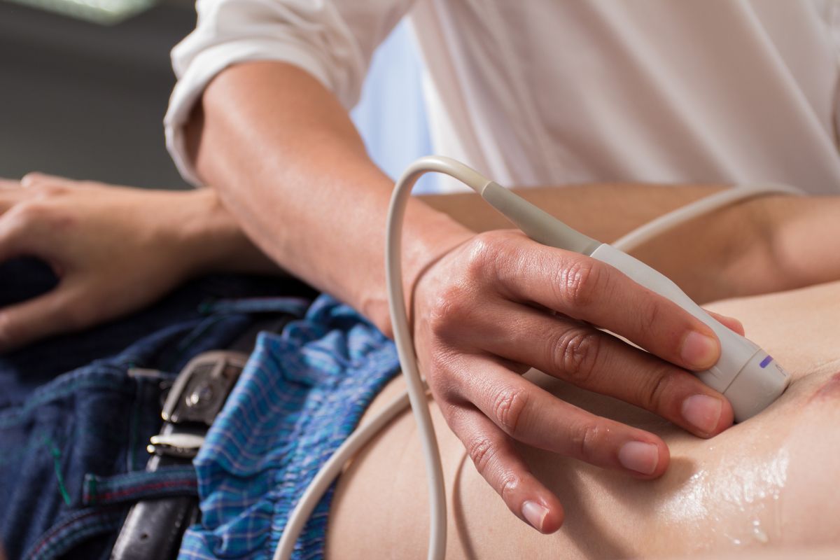
What happens during echo?
During an echocardiography, also called an echo test, a technician or doctor uses a small hand-held device called a transducer to transmit sound waves through the chest to create images of the heart. The sound waves bounce back or echo off the heart and are then translated into images and sounds using a computer.
The echo test is painless and non-invasive. It can help doctors evaluate the size and function of the heart, detect abnormalities such as fluid buildup, blood clots, or heart valve problems, and assess blood flow through the heart.
The test can also help identify any problems with the heart’s muscles, chambers, and valves that may be causing symptoms such as chest pain, shortness of breath, or fatigue.
The entire test usually takes less than an hour, and the results are usually available right away. The doctor will interpret the findings and discuss any necessary treatments or follow-up tests.


